
Paws, Claws and Probes. Oh My!
by C. L. Stambush
A Tale of Ultrasounds in the Animal Kingdom
 The hissing and long, sharp claws unnerved alumna and USI faculty member Dr. Kathy Peak the most.
Mesker Park Zoo & Botanic Garden's Komodo dragon was not a fan of the probe Peak rubbed on her belly either. "Most of the time the animals are sedated or under general anesthesia, but this one was not," said Peak, USI's Clinical Assistant Professor of Radiologic and Imaging Sciences, who holds four degrees from USI: AS'92, BS'11, M'16, D'22. "They just carried her out and plopped her down on a table; she wasn't really having it. She has very long claws and every time I would move the probe around, she would hiss and jerk."
Trained first as a radiologic technologist and then crosstrained in ultrasound, Peak wasn't educated in animal anatomy, and reptile organs weren't remotely familiar. Still, she deftly scanned the unhappy dragon's tummy searching for a reason behind her mood changes.
"She was not acting right, and they were afraid she was egg-bound. That's where they develop eggs, but don't lay their eggs," Peak said. "[The Zoo's staff] wanted me to see if there were any eggs in her that had not passed; to see if that was causing her issue."
Peak has scanned a variety of animals in the six years she's partnered with the Zoo as a community resource. "I've had the opportunity to scan the François langur monkeys, the De Brazza's monkeys, the red-ruff lemurs and the ring-tailed lemurs, the red panda, the clouded leopard, some squirrel monkeys, the jaguar—Beliza—howler monkeys, Fong the sun bear and the Komodo dragon."
USI's relationship with the Zoo began about a decade ago. "My former program chair, Claudine Fairchild, used to go to the Zoo at the request of their veterinarian, Dr. Maria Sprigg, to help out with exams. Dr. Sprigg was a big proponent of being able to utilize community resources that were available to her." Ultrasounds are part of the Mesker Park Zoo's annual wellness checks of its animals, along with physical and dental examinations, X-rays and lab work. "They just like to have some imaging studies done to make sure there is nothing untoward going on with the animals," said Peak. "If we do find something, they could proactively head that off at the pass before it gets worse."
Peak's expertise has been called on for research projects, too, as in the case of the binturong, Vivvy, pregnant with male and female twins: Pretzel and Poppy. "I did head measurements of the babies to help develop growth tables for that species based on head size and hormone levels found in mom's feces."
The Zoo's Health Services Department is equipped with a small examination/surgery room, but many of the Zoo's animals won't fit through the door. In those cases, the medical team goes to them. Beliza, the jaguar who was diagnosed with cancer in 2019, is the largest animal Peak has scanned, and her favorite, noting the big cat's fur is soft and not wiry as she'd expected.
"Since she is large and a more dangerous animal, instead of coming to the veterinary suite, Zoo staff took all of their equipment to her enclosure," Peak said. "They sedated her there with a tranquilizer dart and waited until she could be safely moved. Then they placed her on their examination table and finished the anesthesia process. From there, I did my exam, and they did all of theirs."
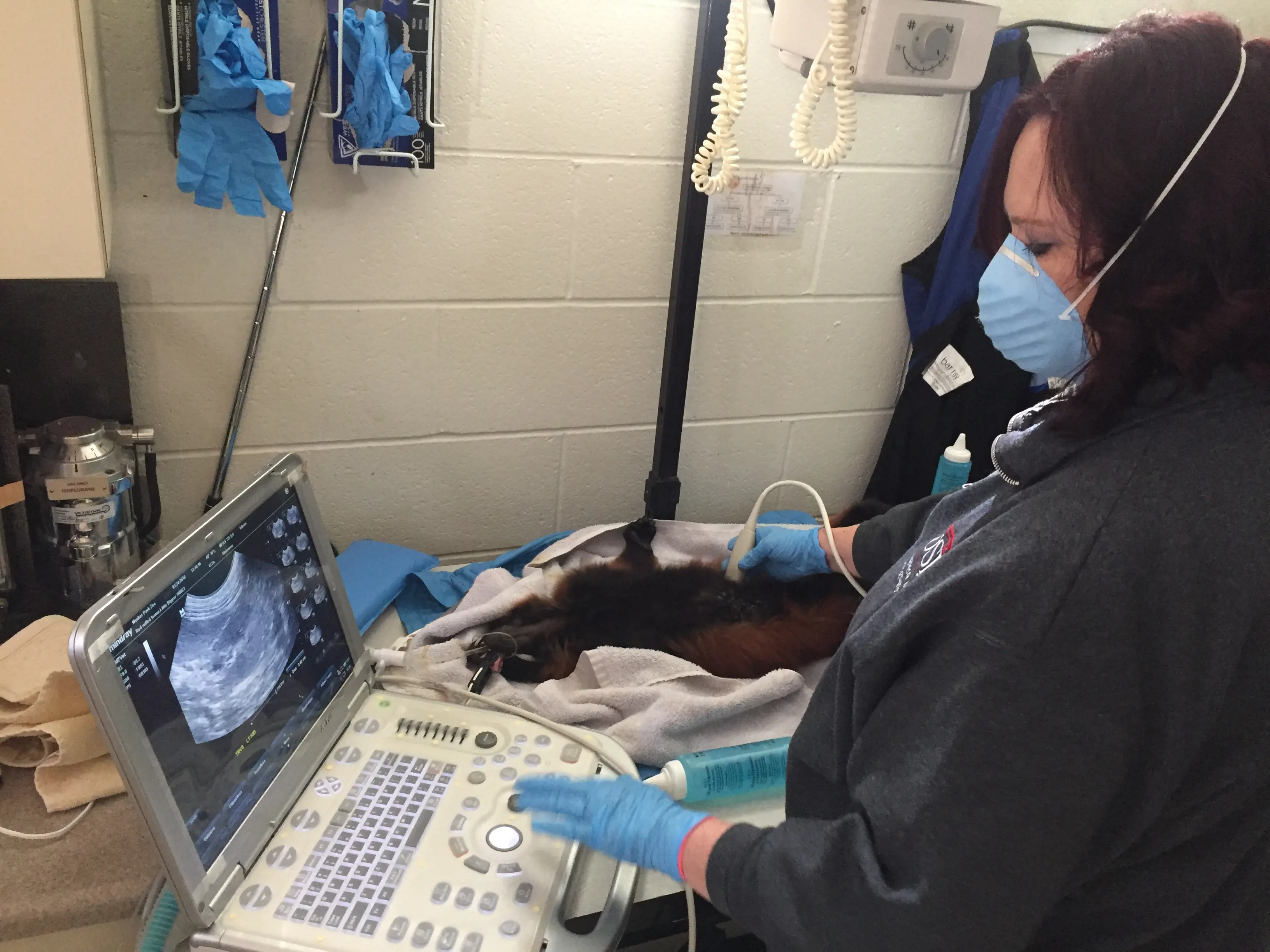
Animal anatomy is different from humans—except for primates, which Peak says are startlingly similar to humans. "Probably the biggest challenge, if they are sedated and not under general anesthesia, is the wiggling and moving around," she said "Even a little bit is like trying to catch a moving target. And the fact that they have fur. With humans we use ultrasound gel and that acts as a couplet between the ultrasound transducer and the patient's skin. But with animals, there is an added layer of fur. You have to use a lot of gel. Sometimes I use [isopropyl] alcohol in spray bottles to mat the fur so the ultrasound waves can penetrate through the fur and skin and into the organs of interest."
Peak started her career as a radiographer and sonographer in a hospital before eventually landing in academia, including eight years as the technical director of a vascular lab. "I loved it and learned so much from the surgeons, but when Claudine Fairchild contacted me about a teaching opportunity at USI, I applied. I've been here for 12 years now."
Ideally, Peak would love to take students with her when she's called to the Zoo. "We have to have the stars aligned where they are not in class and they are not in clinical education, but there has been at least one [time I took] a student with me," she said. "She was able to scan the animal we were looking at that day."
Since her students can't go to the Zoo, Peak brings the Zoo to them. "I always try to update my students on what I've been looking at, show them some pictures, if I'm able to, tell them what kind of things I found, what we were looking for and the purpose of the study," she said. "They always find that very interesting."
Peak's passion for her students is obvious, it's one of the reasons she was awarded the 2023 Faculty Development Award for the College of Nursing and Health Professions. "It's probably the hardest job I've ever had, but hard in a different way. I have to think in a different way now: how to challenge my students; how to convey the information that comes so naturally to me, at this point in my career, to people who are just learning how to do it."
Mentoring and collaborating make-up the rhythm of Peak's career. "I had some really great mentors that I'm still in contact with," she said. "They taught me a lot; I feel like I owe them my career." Now, Peak is training the next generation of professionals. "One of the most rewarding things to me is when someone calls me their mentor. That's one of the highest compliments they can give me."
Medical Imaging Meets Market Demands
Diagnostic medical sonography (DMS), also called ultrasound, has been part of USI's educational offerings for 19 years, but it did not become its own degree program until 2021. Initially, it was offered to radiology students interested in learning ultrasound as a completion program that added a year or two to their studies and housed under the Radiologic and Imaging Sciences Department. "Now when students graduate, their diplomas say Diagnostic Medical Sonography, instead of Radiologic and Imaging Sciences," said Dr. Kathy Peak, Clinical Assistant Professor of Radiologic and Imaging Sciences.
Sonography is the science of using sound waves to create a diagnostic image to investigate an illness, injury or possible treatment of a disease. A sonographer works under the direction of a physician to perform sonographic examinations of many parts of the human body or, in the case of the work Kathy Peak does with Mesker Park Zoo, animal bodies. 
At USI, Peak splits her time teaching in both the DMS and Radiologic Technology (RT) programs. She's excited by the growth happening in RT, too. "With the building renovations in the Health Professions Center, we are getting an entirely new lab and classroom that will include four energized (working) digital X-ray rooms, an energized CT scanner and an MRI simulator closely resembling a working MRI machine."
The new state-of-the-art equipment accompanied by advanced imaging software ensures USI's radiologic and imaging students develop outstanding practical skills through hands-on training and small class sizes. That's probably why since 2008, 100% of graduates seeking employment within the field of imaging sciences were employed within a year of graduation.


